| SpectraBase Spectrum ID |
KlxdQCMepuS |
| Name |
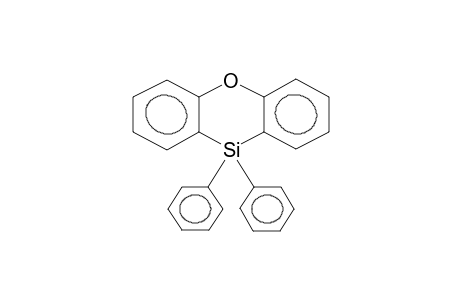
10,10-DIPHENYLPHENOXASILINE |
| Comments |
STRUCTURE DEFINED (A.Y.) |
| Copyright |
Copyright © 2002-2024 Wiley-VCH Verlag GmbH & Co. KGaA. All Rights Reserved. |
| Formula |
C24H18OSi |
| InChI |
InChI=1S/C24H18OSi/c1-3-11-19(12-4-1)26(20-13-5-2-6-14-20)23-17-9-7-15-21(23)25-22-16-8-10-18-24(22)26/h1-18H |
| InChIKey |
QYNNZVFBPRIHES-UHFFFAOYSA-N |
| Instrument Name |
Bruker WM-360 |
| Literature Reference |
V.O.REICHSFELD, S.V.NESTEROVA, N.K.SKVORTSOV, E.L.KUPCHE, E.YA.LUKEVITS (1986)Zhurn.Obsch.Khim.(Russ. Lang.): v.56, N6, 1306-1308. |
| NMR Standard |
TMS |
| Observed nucleus |
29Si |
| Origin |
Chemical Concepts. A Wiley Division. Weinheim, Germany |
| Solvent |
CDCl3 chloroform-d |
