| SpectraBase Spectrum ID |
K46Cij9I4Pb |
| Name |
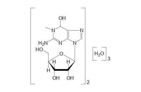
Guanosine, 1.5 H2O |
| Source of Sample |
Calbiochem, EMD Chemicals, Inc., an Affiliate of Merck KGaA, Darmstadt, Germany |
| Catalog Number |
3710 |
| Lot Number |
400603 |
| CAS Registry Number |
118-00-3 |
| Compound Type |
Pure |
| Copyright |
Copyright © 2012-2024 John Wiley & Sons, Inc. All Rights Reserved. |
| Formula |
C11H17N5O5 |
| InChI |
InChI=1S/2C11H17N5O5.3H2O/c2*1-15-9(20)5-8(14-11(15)12)16(3-13-5)10-7(19)6(18)4(2-17)21-10;;;/h2*3-4,6-7,9-10,17-20H,2H2,1H3,(H2,12,14);3*1H2/t2*4-,6-,7-,9?,10-;;;/m11.../s1 |
| InChIKey |
RCTTYQVPXKRJMX-WINRSHHFSA-N |
| Raman Corrections |
Referenced to internal white light source; 180 Degree backscatter |
| Raman Laser Source |
Nd: YAG |
| Raman Laser Wavelength |
1064 |
| Sadtler IR Number |
DW1391 |
| Source of Spectrum |
Forensic Spectral Research |
| Technique |
FT-Raman |
