| SpectraBase Spectrum ID |
Ce26rNGNfel |
| Name |
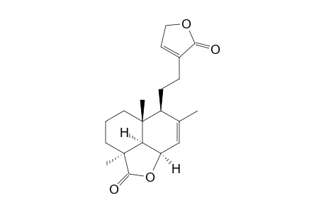
Hypopurin D |
| Appearance |
Colorless prisms |
| Copyright |
Copyright © 2020-2024 John Wiley & Sons, Inc. All Rights Reserved. |
| Formula |
C20H26O4 |
| InChI |
InChI=1S/C20H26O4/c1-12-11-15-16-19(2,8-4-9-20(16,3)18(22)24-15)14(12)6-5-13-7-10-23-17(13)21/h7,11,14-16H,4-6,8-10H2,1-3H3/t14-,15+,16+,19+,20-/m0/s1 |
| InChIKey |
CKFAKQNJAVDAAI-AFJOWOCMSA-N |
| Instrument Name |
Finnigan MAT GCQ/Finnigan MAT 95S |
| Ionization Type |
EI |
| Literature Reference DOI |
10.1021/np0497402 |
| Molecular Weight |
330.424 g/mol |
| Optical Rotation |
[a]25D = +15 (c = 0.2, MeOH) |
| Reported Formula |
C20H26O4 |
| SMILES |
C1C[C@]2([C@@]3([C@](C1)(C(O[C@@]3(C=C([C@@]2(CCC1=CCOC1=O)[H])C)[H])=O)C)[H])C |
| SPLASH |
splash10-01b9-0691000000-543627bb43ab4b772e37 |
| Source of Spectrum |
G4-67-1949-4 |
| Wiley ID |
1881582 |
