| SpectraBase Spectrum ID |
5cGXAdAbWFQ |
| Name |
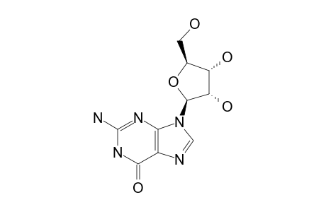
GUANOSINE |
| Copyright |
Copyright © 2016-2024 W. Robien, Inst. of Org. Chem., Univ. of Vienna. All Rights Reserved. |
| Formula |
C10H13N5O5 |
| InChI |
InChI=1S/C10H13N5O5/c11-10-13-7-4(8(19)14-10)12-2-15(7)9-6(18)5(17)3(1-16)20-9/h2-3,5-6,9,16-18H,1H2,(H3,11,13,14,19)/t3-,5-,6-,9-/m0/s1 |
| InChIKey |
NYHBQMYGNKIUIF-GIMIYPNGSA-N |
| Literature Reference Author |
G.REMAUD,X.X.ZHOU,C.J.WELCH,J.CHATTOPADHYAYA |
| Literature Reference Citation |
TETRAHEDRON,42,4057(1986) |
| Literature Reference DOI |
10.1016/S0040-4020(01)87562-1 |
| Solvent |
DMSO;TFA=2EQ |
| Source File Reference |
UWRH493 |
